Reagents > Histone H3 Mouse Monoclonal Antibody for Normalization

The Histone H3 primary antibody can be used as an internal loading control for normalization and is particularly effective when detecting target proteins in nuclear extracts.
The expression of Histone H3, or any housekeeping protein (HKP), should be validated to ensure that its expression does not change under experimental conditions.
Once validated, Histone H3 primary antibodies can be used for the detection of Histone H3 when performing multiplex Western blot detection.
Detect Histone H3 Mouse Monoclonal Antibody with IRDye® Goat anti-Mouse or IRDye Donkey anti-Mouse secondary antibodies.
组蛋白 H3 一抗可用作标准化的内部上样对照,在检测核提取物中的靶蛋白时特别有效。
应验证组蛋白 H3 或任何管家蛋白 (HKP) 的表达,以确保其表达在实验条件下不会改变。
经验证后,在进行多重蛋白质印迹检测时,组蛋白 H3 一抗可用于检测组蛋白 H3。
使用 IRDye® 山羊抗小鼠或 IRDye 驴抗小鼠二抗检测组蛋白 H3 小鼠单克隆抗体。
Other options for housekeeping protein normalization
Reactivity and Specificity
Histone H3 antibody is supplied in 10 mM HEPES (pH 7.5), 150 mM NaCl, 100 µg/mL BSA, 50% glycerol, and <0.02% sodium azide.
组蛋白 H3 抗体以 10 mM HEPES (pH 7.5)、150 mM NaCl、100 µg/mL BSA、50% 甘油和 <0.02% 叠氮化钠的形式提供。
Do not aliquot the antibody.
| Properties | Histone H3 Mouse Monoclonal Antibody (P/N 926-42218) |
|---|---|
| Species Cross-Reactivity | Human, mouse, rat, monkey |
| Target Molecular Weight | 17 kDa |
| Isotype | Mouse IgG3 |
| Specificity/Sensitivity | Detects endogenous levels of total Histone H3 protein |
| Immunogen | A peptide that is specific to the carboxy terminus of human histone H3 protein |
| Tested Applications | Western blot (WB), Immunohistochemistry (IHC), Immunofluorescence (IF), Flow Cytometry (F), Chromatin Immunoprecipitation (CHIP)
蛋白质印迹 (WB)、免疫组织化学 (IHC)、免疫荧光 (IF)、流式细胞术 (F)、染色质免疫沉淀 (CHIP) |
RRID
- P/N 926-42218: RRID AB_2814903
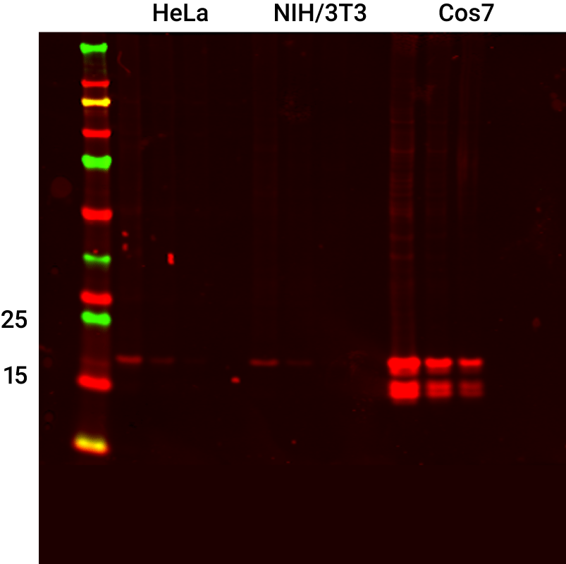
Detection of Histone H3 Mouse Monoclonal Antibody in HeLa, NIH/3T3, and COS-7 Lysates